


Increasingly, researchers are using technical CT scanners to examine instruments for construction secrets to woodworm and previous repairs. Rudolf Hopfner presents a guide to the technology and the microscopic details it can reveal
This is an extract from a longer article in The Strad’s January 2018 issue. To read further and view more photos, download now on desktop computer or via the The Strad App, or buy the print edition
In the past few years, high-resolution CT scanning has become a standard procedure in the testing of materials, as well as in many other fields of engineering. In tandem, the technique has become increasingly employed in the documentation of items representing our cultural heritage – especially stringed instruments. As curator of the collection of historic musical instruments at Vienna’s Kunsthistorisches Museum, I have a 15-year acquaintance with computed tomography, from when we had the opportunity to scan a newly acquired Stainer violin in 2003. The scan (figure 1, see below) was facilitated by the University of Vienna’s Department for Veterinary Medicine and the result was ambiguous. For the first time we could scrutinise the interior and learn some details of the instrument’s construction; but it was disappointing because the low resolution of the machine delivered only blurred images.

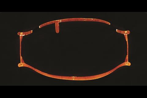
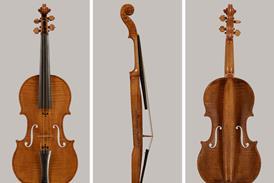
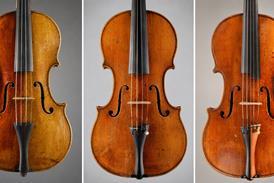
Ten years later, in 2013, I got in touch with Gerhard Weber, head of the Vienna micro-CT (μCT) lab. This institution runs a high-resolution technical CT scanner with a gantry big enough to accommodate a violin or a small viola. The ‘patient’ once again was the Stainer violin from our collection. The results of the μCT scan were a revelation (figure 2, see above). Tiny details such as the annual rings of densely grown spruce or the shape of the pinholes in the back of the instrument, typical of Stainer’s work, became visible.
What is the difference between medical and technical CT scanners? For obvious reasons, medical scanners are optimised for short acquisition times and low-radiation doses. The machine settings are customised for certain standard examination scenarios. On the contrary, technical CT scanners aim for high resolution and high flexibility in the adjustment of all scan parameters. Exposure and acquisition time, as well as radiation dose, usually are of less importance. Figures 1 and 2 depict the same axial section through the body of the Stainer violin. The size of the voxels, which defines the maximum resolution, is 0.45 x 0.45 x 2mm for the medical scan and 0.1 x 0.1 x 0.1mm for the μCT scan. Of course, this increase in resolution comes at a cost. The acquisition time for a μCT scan of a violin body can amount to eleven hours. The storage space for the Stainer violin is around 150MB for the medical scan, whereas the μCT scan required 12GB.
To see the full article, download The Strad’s January 2018 issue on desktop computer or via the The Strad App, or buy the print edition


1 Readers' comment